

Overview & Features
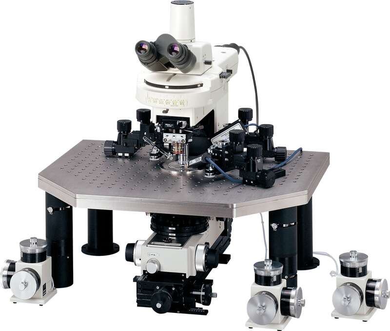
The ECLIPSE FN1, developed especially for electrophysiological research, features enhanced operability to facilitate patch-clamp experiments. Advantages offered by the FN1 include slim body, streamlined structure, improved electrode placement, long working distance, and greater noise reduction. A deeper area of the specimen can be observed clearly with infrared (IR) light.
Features
The ECLIPSE FN1, developed especially for electrophysiological research, features enhanced operability to facilitate patch-clamp experiments. Advantages offered by the FN1 include slim body, streamlined structure, improved electrode placement, long working distance, and greater noise reduction. A deeper area of the specimen can be observed clearly with infrared (IR) light.
One lens covers from low to high magnifications
The high NA, long working distance 16x objective (NA 0.8, WD 3.0) allows observation at broad magnification range from 5.6x to 64x when combined with an optional variable magnification double port. This objective enables observation from a low magnification wide field of view (up to 2.0mm) to a high magnification high-resolution field without changing objectives.


Image deeper areas with ultimate clarity with IR-DIC
Axial chromatic aberration in the near-infrared region (up to 850nm) has been corrected in CFI Apochromat NIR 40X/60X W for clearly observing the minute structure of thick specimens. The CFI Plan 100XC W (NA 1.1, W.D. 2.5) is the world’s first water dipping lens with depth-induced aberration correction. Because of its special correction ring, this lens can correct spherical aberration induced by imaging deep in tissue, or by working at physiological temperatures. Consequently, it is ideal for IR-DIC imaging, confocal applications, and multiphoton imaging. Alternating between visible and infrared wavelengths, as well as different illumination techniques (DIC to Oblique Light) is carried out simply by rotating the respective turrets.

Easy electrode placement
Access of microelectrodes to the specimen is easy as the objectives have a long W.D. of 2.5-3.5mm and broad approach angle up to 45°.

Safe, easy objective changeover
Objectives mounted on the sliding nosepiece can be raised when switching magnifications. This prevents the objective from colliding with the manipulator or the chamber. The retraction distance is 15mm, so even a thick glass dish is protected. The lens top can be easily dipped (approximately 1mm) into the bath solution by using the lever to eliminate the risk of specimen disturbance.

Operational within your reach
The focus knob and field diaphragm ring are located on the front part of the microscope base, and there are no cumbersome outside belts; so operation is easy when using a fixed stage. An optional remote handle is also available to enable operation from outside the cage.

Enhanced noise reduction
Electrical noise has been successfully reduced by utilizing fiber illumination to bring light into the system from outside the cage and by connecting ground pins to all main parts of the microscope. Vibration noise has also been reduced by undertaking critical measurement and simulation analysis to improve microscope rigidity.

Responsive to a broad range of experimental needs
By inserting a spacer between the body and the arm, you can raise the microscope height 10-30mm. This facilitates large specimen observation. Moreover, the condenser, sub-stage and turret can be removed entirely from the microscope to allow for more free space, depending on the purpose of the experiments.

Specifications
| Eclipse FN1 | |
|---|---|
| Optical system | CFI60 and CFI75 infinity optical system |
| Main body | I-shaped, external power supply |
| Focusing | Via nosepiece up/down movement Manual coaxial coarse/fine focus knobs (on both sides) |
| Nosepiece | FN-S2N Sliding Nosepiece (for CFI60 objectives) Front/back 2-position; DIC prism attachable FN-MN-N Single Objective Holder (for CFI75 objective) 1-position; DIC prism attachable |
| Objectives | CFI Plan 4X, NA: 0.10, W.D.: 30.0 (*1) CFI Plan Fluor 10X W, NA: 0.30, W.D.: 3.5 CFI75 LWD 16X W, NA: 0.80, W.D.: 3.0 CFI Apochromat NIR 40X W, NA: 0.80, W.D.: 3.5 CFI Apochromat NIR 60X W, NA: 1.00, W.D.: 2.8 CFI Plan 100XC W, NA: 1.10, W.D.: 2.5, with correction ring |
| LWD condenser | Universal turret type NA: 0.78, W.D.: 7.2mm DIC and Oblique Light observations possible |
| Eyepiece | 10x, F.O.V.: 22 UW10x, F.O.V.: 25 |
| Eyepiece tubes | C-TE2 Ergonomic Binocular Tube (Bino 100%, Bino : DSC port = 50 : 50) (DSC port cannot be used with variable magnification double port) C-TF Trinocular Tube F (Bino : Photo = 100 : 0, 0 : 100) C-TT Trinocular Tube T (Bino : Photo = 100 : 0, 20 : 80, 0 : 100) LV-TI3 Trinocular Tube ESD (Bino : Photo = 100 : 0, 0 : 100) LV-TT2 Tilting Trinocular Tube (Bino : Photo = 100 : 0, 20 : 80, 0 : 100) |
| Stage | FN-3PS2 FN1 Rectangular Stage (3-plate mechanical stage) Stroke: 30mm (X, Y) |
| Light source | Intensilight HG Precentered Fiber Illuminator: 130W long-life mercury lamp Hg Lamphouse: 100W mercury lamp FN-LH Precentered Lamphouse: 12V-100W long-life halogen lamp |
| Operating conditions | Temperature: +10ºC to +40ºC Humidity: 85% RH max. (no condensation) |
| Weight (main body) | Approx. 12kg |
*1 Auxiliary lens is required.